
News and Announcements
Hip Arthroscopy: The Ultimate Patient Guide – Answers to All Key Questions
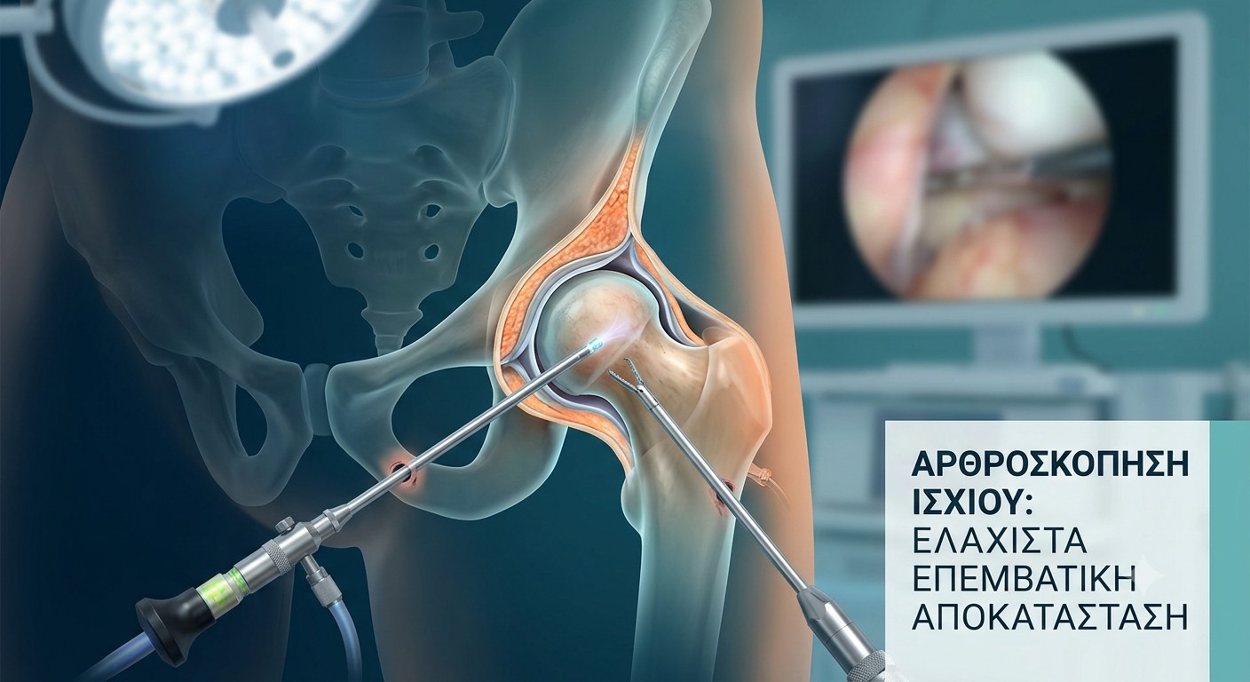
Hip Arthroscopy: Diagnostic Overview, Recovery Timeline, and Surgical FAQs
Hip arthroscopy is a modern, minimally invasive surgical technique. Instead of large traditional incisions, the surgeon uses a miniaturized camera (the arthroscope) and specialized instruments inserted through 2 to 3 small portals measuring just a few millimeters.
High-definition visual guidance allows simultaneous diagnosis and treatment of internal hip joint pathology—such as repairing a labral tear or reshaping dynamic bone deformities (Cam and Pincer lesions)—without disrupting the surrounding muscular architecture.
1. Is hip arthroscopy a permanent solution?
Yes. When performed before the onset of advanced osteoarthritis, joint reshaping provides a definitive correction. Eliminating structural impingement (Cam/Pincer morphology) removes the mechanical cause of friction, protecting the articular cartilage from future degradation.
2. Is the procedure indicated if osteoarthritis is already present?
Hip arthroscopy yields optimal results in early to moderate stages of joint wear where articular cartilage structure remains intact. In advanced (end-stage) osteoarthritis with extensive cartilage loss, arthroscopy cannot regenerate the joint surface. In these cases, Robotic MAKO Total Hip Arthroplasty is the gold standard for full functional restoration.
3. What is the expected timeline for returning to work?
-
Desk jobs: 7–10 days post-op, as sedentary activity places minimal mechanical stress on the healing joint.
-
Manual labor / Physical roles: 2–3 months, depending on lifting requirements, prolonged standing, and physical demand.
4. When is it safe to resume driving?
Driving is generally permitted at 2 weeks post-operation, provided you have discontinued assistive devices (crutches), regained sufficient lower-limb motor control, and can apply immediate, full emergency braking force without pain.
5. What are the potential risks and complications?
Serious complications occur in less than 5% of cases. The most frequent transient side effect is mild numbness (hypoesthesia) on the dorsum of the foot, groin, or perineal area. This is caused by the temporary limb traction required during surgery to create joint clearance; it is fully reversible and resolves within days.
6. Can a labral tear heal on its own? What is the current standard of care?
No. The acetabular labrum has poor vascularity and limited self-healing capacity. Conservative treatment may temporarily reduce inflammation, but it cannot restore anatomical integrity.
Current surgical standards prioritize arthroscopic labral repair using bioabsorbable anchors over simple debridement. Preserving the labrum maintains the joint's fluid seal (suction seal mechanism), which stabilizes hydrostatic pressure and protects against premature cartilage degradation. If the tissue is irreversibly damaged, labral reconstruction using a graft is performed.
7. What does the post-operative rehabilitation protocol look like?
-
Hospital Discharge: Same day or the day following surgery.
-
Weight-Bearing: Partial weight-bearing with crutches for the first 6 weeks to protect labral repairs.
-
Physical Therapy: Begins immediately to restore range of motion and progressive pericapsular strength.
-
Daily Activities: Full routine mobility within 1–2 months.
-
Sports & Athletics: Full return to high-impact sports in 4–6 months.
8. How do I choose between Arthroscopy/Biologics and MAKO Robotic Surgery?
| Treatment Option | Patient Profile & Indications | Clinical Objective |
| Arthroscopy & Biologics (PRP / Hyaluronic Acid) | Younger, active patients with mechanical impingement (Cam/Pincer), labral tears, or early-stage cartilage wear. | Preserve the native joint, correct mechanics, and delay/prevent osteoarthritic progression. |
| Robotic MAKO Arthroplasty | Patients with severe, end-stage osteoarthritis and complete cartilage loss ("bone-on-bone"). | Provide a definitive total joint replacement, eliminate chronic pain, and restore mobility. |
9. What are the primary warning signs of a hip issue?
-
Deep groin (inguinal) pain that may radiate to the thigh or buttock.
-
Pain exacerbated by prolonged sitting (e.g., driving, desk work) or rotational movements.
-
Mechanical symptoms such as clicking, catching, or joint locking.
-
Progressive stiffness limiting daily tasks (e.g., difficulty putting on socks or shoes).
10. Where are these procedures performed?
Hip arthroscopy requires advanced surgical equipment and specialized training. These procedures are performed in dedicated medical centers and modern private hospitals in major metropolitan areas equipped with high-definition arthroscopic technology.
Hip arthroscopy addresses the anatomical root cause of mechanical joint pain rather than merely managing symptoms. By restoring structural integrity early, it offers patients a clear pathway back to an active, high-quality lifestyle.


